| |
|
|
The most common echocardiographic views |

|

|
|
Thursday, 05 February 2004 |
Click on each figure to increase it - If the enlarging does not function, check in your browser's option that 'javascript' is well activated.If echocardiography is to be used in intensive care, there is a need for (i) an intensivist trained in general echocardiography but also in intensive care echocardiography, (ii) an echocardiographer dedicated to the intensive care unit, (iii) a multiplane transesophageal probe. Unless there is a diagnostic emergency, we reserve transesophageal echocardiography (TEE) for sedated and mechanically ventilated patients, bearing in mind the difficulty of performing such an examination in a spontaneously breathing patient presenting, for example, acute respiratory failure. Echocardiography is used for diagnostic purposes (endocarditis, valve disease, dissection of the aorta, aortic trauma, pericardial effusion, pulmonary embolism, cardiomyopathy…) and also for therapeutic reasons, such as evaluation of central blood volume, cardiac function, and impact of ventilation on cardiac function, for decision-making regarding volume expansion, infusion of inotropic agents, changes to ventilator settings, inhalation of NO…
The transthoracic route is often difficult in intensive care, because of the examination conditions (supine patient, patient suffering pain, patient with tachypnea, assisted ventilation), but allows rapid patient management. The following principal views should be obtained where possible:
(i) An apical four-chamber view (Film 1). This enables assessment of the size of the cardiac chambers, detection of any mitral and aortic valve disease, Doppler echocardiography of the mitral annulus and of the LV outflow tract of the left ventricle (calculation of cardiac flow).
(ii) A two-dimensional, long-axis parasternal view (Film 2) completed using motion mode (Film 3). This enables measurement of the size of the two ventricles, calculation of the left ventricular ejection fraction from estimated volumes (Teicholz), and examination for paradoxical septal movement.
(iii) A short-axis parasternal view (Film 4) is the most sensitive route for detection of paradoxical movement of the septum, and enables measurement of the LV fractional area contraction, close to its ejection fraction. From this view one can also obtain a view of the vessels at the base of the heart, thus enabling Doppler echocardiography of the pulmonary artery, which is very often informative in pulmonary arterial hypertension (Film 5).
(iv) Finally, the subcostal view is important as it visualizes the inferior vena cava and its variations during ventilation (Film 6), and because it is often available and of good quality in a mechanically ventilated patient. The transesophageal route, however, remains the route of choice for echocardiography in a sedated, mechanically ventilated patient. Most of the different effects have their counterpart in the transthoracic route. It is essential to display the airway pressure signal on the echocardiograph screen throughout the hemodynamic study, so as to pinpoint cardiac events in the respiratory cycle.
The long-axis view of the left ventricle (Film 7) is close to the transthoracic, apical, four-chamber view. It visualizes the four cardiac chambers, albeit truncating the size of the two ventricles by about 12 to 15%. This view can be used to record Doppler flow at the mitral annulus (Film 8).
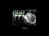 Figure 3 : TEE long-axis view of the left ventricle in a patient ventilated because of cardiogenic acute pulmonary edema related to myocarditis. Pulsed Doppler at the mitral annulus. LV: left ventricle, RV: right ventricle, LA: left atrium. E: rapid protodiastolic filling of the LV. A: end-diastolic filling of the LV (atrial systole). Restricted mitral flow (E/A > 2, deceleration time of the brief E wave) suggestive of high end-diastolic pressure of the left ventricle Figure 3 : TEE long-axis view of the left ventricle in a patient ventilated because of cardiogenic acute pulmonary edema related to myocarditis. Pulsed Doppler at the mitral annulus. LV: left ventricle, RV: right ventricle, LA: left atrium. E: rapid protodiastolic filling of the LV. A: end-diastolic filling of the LV (atrial systole). Restricted mitral flow (E/A > 2, deceleration time of the brief E wave) suggestive of high end-diastolic pressure of the left ventricle
|
The short-axis view of the left ventricle by the transgastric route must pass by the cords of the mitral valve (Film 9). It is obtained by pushing the probe in the stomach and then tilting it upwards slightly. As with the transthoracic route, it can be used to evaluate the contractile function of the LV and to visualize any paradoxical movement of the septum. By appropriate positioning of the probe, it is possible to scan the left ventricle from the apex towards the base. By tilting the multiplane probe by 120°, this view unveils the LV outflow tract of the left ventricle, enabling pulsed Doppler for calculation of cardiac flow (Film 10). In certain patients it is possible, again by the transgastric route, to visualize the outflow tract of the right ventricle and the pulmonary artery so as to obtain a Doppler signal (Film 11).
By withdrawing the probe from the long-axis view while tilting it upwards, it is possible to unveil, among other things the left atrium and the left superior pulmonary vein (Film 12).  Figure 4 : TEE view passing through the left superior pulmonary vein in the same patient as in figure 3. S: systolic wave, D: diastolic wave. a: retrograde wave concomitant with atrial systole. The reverse view of the flow of left atrial filling (small systolic wave, large diastolic wave), and the presence of a significant A wave, are suggestive of high LV end-diastolic pressure Figure 4 : TEE view passing through the left superior pulmonary vein in the same patient as in figure 3. S: systolic wave, D: diastolic wave. a: retrograde wave concomitant with atrial systole. The reverse view of the flow of left atrial filling (small systolic wave, large diastolic wave), and the presence of a significant A wave, are suggestive of high LV end-diastolic pressure |
By continuing to withdraw the probe to about 30 centimeters from the esophageal opening, one obtains an extremely useful view which allows Doppler flow recording in the pulmonary artery downstream of the sigmoid arteries (Film 13). It also unveils the superior vena cava and allows longitudinal-view study (at 90°) of its variations during the respiratory cycle (Film 14). Finally, it visualizes the trunk of the pulmonary artery, the right pulmonary artery, and the beginning of the left pulmonary artery when seeking a thrombus. 
|
|
|
| |
|


)
)